Regulation of metabolism and immunity
Nuclear receptors
 We are investigating molecular biology and pathophysiology of nuclear receptors, particularly lipid-sensing receptors such as liver X receptor, FXR and VDR, and other metabolic regulators to elucidate mechanisms of human diseases and to develop new therapies (Makishima et al., J Pharmacol Sci 2005).
VDR is a dual functional receptor for vitamin D and LCA
The oxysterol receptors, liver X receptor (LXR) alpha and LXRbeta, regulate cholesterol metabolism and the bile acid receptor, farnesoid X receptor (FXR), controls bile acid metabolism (Makishima et al., Science 1999; Lu and Makishima et al, Mol Cell 2002). Vitamin D receptor (VDR) has been identified as a receptor for 1,25-dihydroxyvitamin D3 (1,25(OH)2D3). VDR also acts as a bile acid receptor (Makishima et al., Science 2002).
We are investigating mechanisms of dual functions of VDR.
- A bile acid lithocholic acid (LCA) binds to the ligand-binding pocket of VDR differently from 1,25(OH)2D3 (Adachi et al., Mol Endocrinol 2004).
- LCA acetate is a potent VDR ligand, which can induce differentiation of myeloid leukemia cells (Adachi et al., J Lipid Res 2005).
- LCA acetate and LCA propionate activate tissue VDR without inducing hypercalcemia (Ishizawa et al., J Lipid Res 2008).
- 1,25(OH)2D3 and LCA induce selective VDR-RXR-cofactor interaction (Choi et al.,Mol Pharmacol 2011; Chuma et al., Biol Pharm Bull 2012).
- Development of tissue- or cell type-selective vitamin D derivatives (Inaba et al., Mol Pharmacol 2007; Kudo et al., J Med Chem 2014; Watarai et al., J Med Chem 2015; Otero et al., J Med Chem 2018; Maekawa et al., Biomolecules 2023).
- p38alpha and GADD45A are involved in 1,25(OH)2D3-induced TRPV6 expression in intestinal cells (Ishizawa et al., Steroid Biochem Mol Biol 2017).
- Zinc signaling selectively inhibits 1,25(OH)2D3-induced E-cadherin gene expression in intestinal cells (Ishizawa et al., Anticancer Res 2021).
VDR and bile acid metabolism
VDR is a regulator of bile acid metabolism and regulates pathogenesis of cholestasis.
- Vitamin D treatment enhances bile acid metabolism, particularly urinary excretion (Nishida et al., Drug Metab Disp 2009).
- VItamin D treatment represses inflammation in mice with bile duct ligation (Ogura et al., J Pharmacol Exp Ther 2009).
- Inflammatory responses are repressed in the intestine of VDR knockout mice (Ishizawa et al., PLoS ONE 2012).
- Lithocholic acid activates VDR selectively in the lower intestine (Ishizawa et al., Int J Mol Sci 2018).
- Fecal bile acid excretion is decreased in VDR knockout mice (Nishida et al., J Nutr Sci Vitaminol 2020).
- Lithocholic acid suppresses colitis in mice in a VDR-dependent manner (Kubota et al., Int J Mol Sci 2023).
Novel LXR ligands
LXR regulates inflamation, immunity, cell growth and apoptosis as well as lipid metabolism.
- Oxysterol derivatives are function-selective LXR ligands (Kaneko et al., J Biol Chem 2003).
- LXR activation supresses beta-catenin signaling (Uno et al., Biochem Pharmacol 2009).
- Metabolites of 7-dehydrocholesterol by CYP27A1 are selective LXR modulators (Endo-Umeda et al., J Steroid Biochem Mol Biol 2014).
- 1alpha-Hydoxy derivatives of 7-dehydrocholesterol are LXR ligands that potently suppress inflammation (Endo-Umeda et al., Steroid Biochem Mol Biol 2017).
Nuclear receptors regulate immunity in the liver and intestine
Hepatic immune cells regulate both metabolism and inflammation/immunity. Both LXR and VDR are expressed in hepatic nonparenchymal cells, such as Kupffer cells/macrophages. However, their cell-specific functions in hepatic immunity remain uncertain. We are investigating the roles of LXR and VDR in regulation of metabolism and immunity in the liver and intestine in animal models using LXR or VDR knockout mice.
- LXR regulates F4/80+CD11b+ macrophage population and inflammation in the liver (Endo-Umeda et al., Sci Rep 2018; reviewed in Endo-Umeda and Makishima. Int J Mol Sci 2019).
- LXRalpha deficiency promotes the progression of NASH by cholesterol accumulation and macrophage activation in the liver (Endo-Umeda et al., Endocrinology 2018; reviewed in Endo-Umeda and Makishima. Int J Mol Sci 2019).
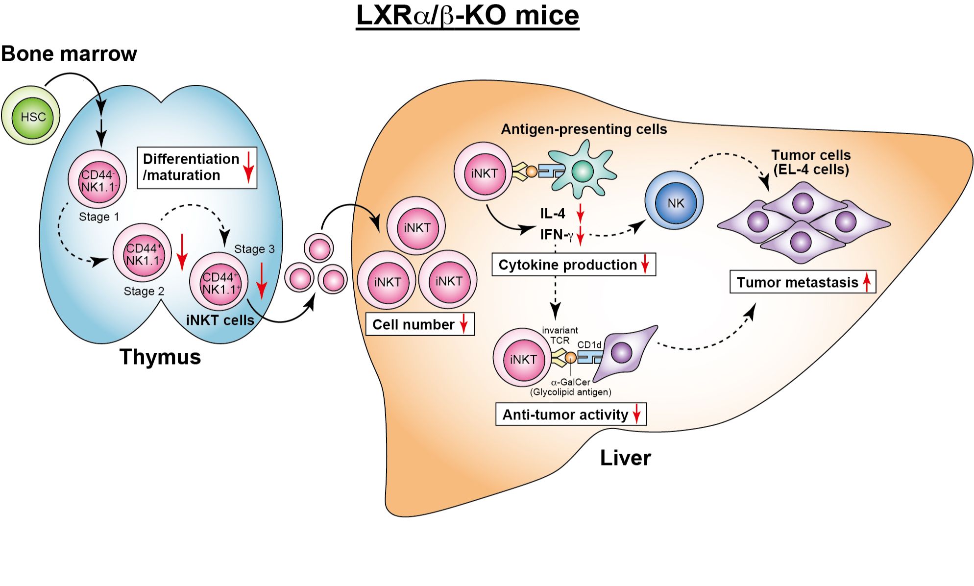
- Natural killer T cells and hepatic antitumor activity are diminished in LXR-deficient mice (Endo-Umeda et al., Sci Rep 2021).
- Concanavalin A-induced hepatitis is attenuated by reducing ROS production by Kupffer cells in VDR-knockout mice (Umeda et al., J Leukoc Biol 2019).
UnSUMOylated PPARgamma
PPARgamma is a receptor for fatty acids, as shown above, and activated upon fatty acid binding. Conversely, its activity is repressed by post-translational modifications such as SUMO conjugation (sumoylation). The unsumoylated PPARgamma mice, which carry PPARgamma with the sumoylation site Lys107 replaced by Arg for blocking its sumoylation, were generated to elucidate the physiological significance. Even after high-fat diet feeding, unsumoylated PPARgamma mice maintained higher glucose tolerance, and exhibited elevated gene expression for energy metabolism and reduced inflammatory gene expression in their adipose tissues, compared with their wild-type counterpart (Katafuchi et al. Proc Natl Acad Sci USA 2018).
FGF15 and C-type natriuretic peptide (CNP)
FGF15: A hormone that regulates bile acid secretion
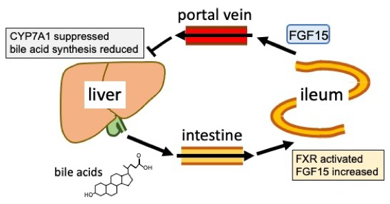
Fibroblast growth factor 15 (FGF15) is one of ileal hormones in rodents, and its ortholog in human is called FGF19. The production of FGF15 in the ileum is increased upon stimulation with bile acids, and the increase is due to activation of the bile acid receptor FXR. Based on the study using FGF15-KO and beta-Klotho-KO mice, the secreted FGF15 travels through the portal vein and stimulate FGFR4/beta-Klotho receptor complex in the liver, which is followed by repressed expression of CYP7A1, the rate-limiting enzyme in bile acid biosynthesis. This study clarified that FGF15 is a feed-back mediator to shut off bile acid biosynthesis shortly after FXR in the ileum detects significant amount of bile acids (Katafuchi et al. Cell Metab 2015).
CNP-induced adipogenesis
Elucidation of underlying mechanism in adipogenesis is generally considered quite important for development of a novel intervention for obesity. One of preadipocyte cell lines 3T3-L1, which undergoes adipogenesis under treatment with an adipogenic medium containing insulin, dexamethasone, a synthetic glucocorticoid, and IBMX, a phosphodiesterase inhibitor for maintaining intracellular cAMP and cGMP levels, and is accepted as a great model to study how adipogenesis occurs. We stimulated 3T3-L1 cells with an adipogenic medium containing insulin, dexamethasone and C-type natriuretic peptide (CNP), a peptide hormone increasing intracellular cGMP production to replace the function of IBMX, and found that CNP strongly induced adipogenesis (Katafuchi et al. Peptides 2010).
AHR
Benzo[a]pyrene (BaP) acitivates aryl hydrocarbon receptor (AHR) and AHR induces expression of CYP1 family enzymes. CYP1 enzymes have been thought to convert BaP to toxic metabolites, a mechanism called metabolic activation. Studies using CYP1 knockout mice show that CYP1 enzymes, particularly CYP1A1, are involved in detoxification of BaP rather than metabolic activation (Uno et al., Mol Pharmacol 2004; Uno et al., Mol Pharmacol 2006).
- CYP1A1 and CYP1B1 metabolite BaP to inactive compounds for AHR and CYP1A1 and CYP1A2 repress BaP-induced DNA adduct formation (Endo et al., Toxicol Appl Pharmacol 2008).
- AHR activation enhances vitamin D inactivation (Matsunawa et al., Toxicol Sci 2009).
- AHR activation exagerates cholestatic liver injury and CYP1A1 plays a role in inhibiting the pathogenesis (Ozeki et al., Toxicology 2011).
- BaP induces arteriosclerosis and CYP1A1 suppresses this pathogenesis (Uno et al., Toxicology 2014).
- BaP aggravates non-alcoholic fatty liver, but this effect is suppressed by CYP1A1 (Uno et al., Food Cheml Toxicol 2018).
- Diallyl trisulfide, a component of garlic essential oil, enhances BaP-induced expression of CYP1A1 and CYP1B1, associated with increased BaP metabolic activation (Uno et al., Anticancer Res 2019).
- BaP suppresses colitis in mice without the toxicity associated with metabolic activation (Adachi et al., Chem Biol Interact 2022).
Transcriptional coregulator ESS2 (DGCR14)
Transcription factors, such as nuclear receptors, regulate their target genes mRNA expression by interacting with proteins, called transcriptional coregulators. Transcriptional coregulators include histone modifying enzymes (acetylation, methylation, and phosphorylation, etc.), chromatin remodeling factors that change chromosome structure, and mediators that mediate interactions with RNA polymerase.
The ESS2 (also called as DGCR14) gene was identified as a deletion gene in 22q11 gene deficiency syndromes such as DiGeroge syndrome and we identified ESS2 as a transcriptional coactivator for RORgamma/gamma-t in T cells (Takada, Mol Cell Biol 2015). Because ESS2 does not have known functional domains and conventional Ess2 knockout mice is early embryonic lethal, we analyzed the functions of ESS2 and found following results.
ESS2 enhances the transcriptional activities of RORgamma/gamma-t and regulates Th17 cell differentiation by associating with the chromatin remodeling factor BAZ1B and the protein kinase RSK2 (Takada, Mol Cell Biol 2015).
BI-D1870, which is an inhibitor for pan-RSKs, suppresses Th17 cell differentiation and ameliorates experimental autoimmune encephalomyelitis in mice (Takada et al., Immunobiology 2016).
ESS2 interacts with RORs at the N-terminal domain and with spliceosomes at the C-terminal domain. ESS2 knockdown reduced the interaction of RORs with lncRNA (Takada I et al., Biochem Biophys Res Commun 2018).
Now, we are studying the molecular mechanism of ESS2 in vivo using newly generated conditional Ess2 knockout mice and studying the effect of RSK inhibitors to autoimmune disease for the application of therapy.
Hepatitis virus, cancer and omics research
Omics research on hepatitis virus infection and cancer
Disease omics is to perform the comprehensive analysis of gene expression and protein profiles in various disease stages followed by comparison; For example, genomics leads to find the DNA alterations specific for disease. Transcriptomics leads to find the RNA expression changes specific for disease. Proteomics leads to find the protein expression changes specific for disease. These results are useful to elucidate the molecular mechanism of disease and to develop the molecular diagnosis and molecular target therapy for disease.
Molecular biology of hepatitis C virus offense and host defense in human liver
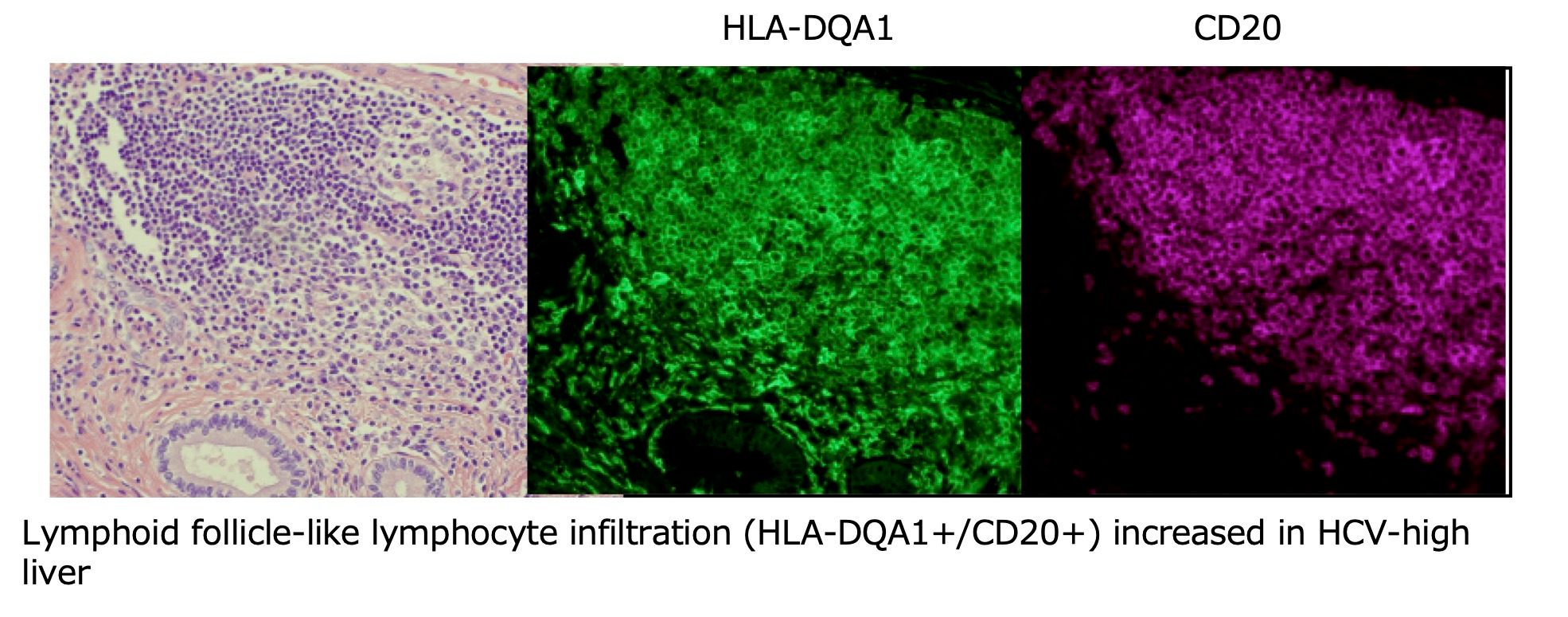
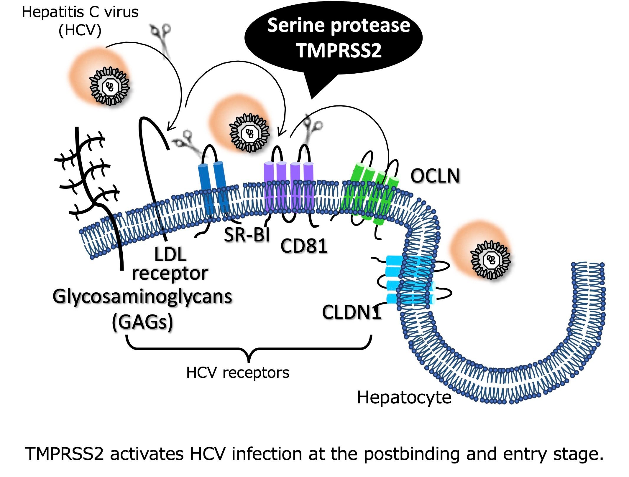
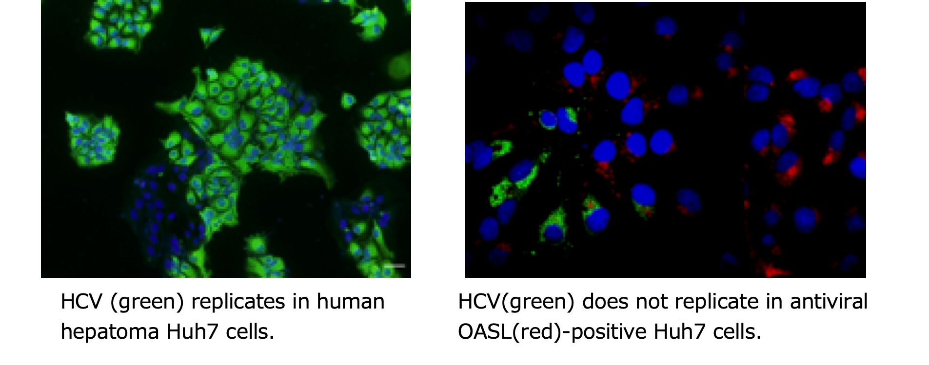
Transcriptomics of liver RNA from two groups of hepatitis C virus (HCV) loads, high and low, revealed 26 genes up-regulated in the HCV-high group (Esumi et al., Hepatology 2015); Of which, HLA-DQA1 indicates the increase of antigen presenting cells in the HCV-high group (Ishibashi et al., Arch Virol 2018). OASL functions to inhibit the HCV replication in vitro (Ishibashi et al., Biochem Biophys Res Commun 2010). Transmembrane serine protease TMPRSS2 functions to activate the HCV infection in vitro (Esumi et al., Hepatology 2015). Liver sinusoidal endothelial cell-specific CLEC4M functions as a receptor for transinfection of HCV in vitro (Ishibashi et al., Arch Virol 2014).
Recurrence markers of human hepatocellular carcinoma from liver tissues
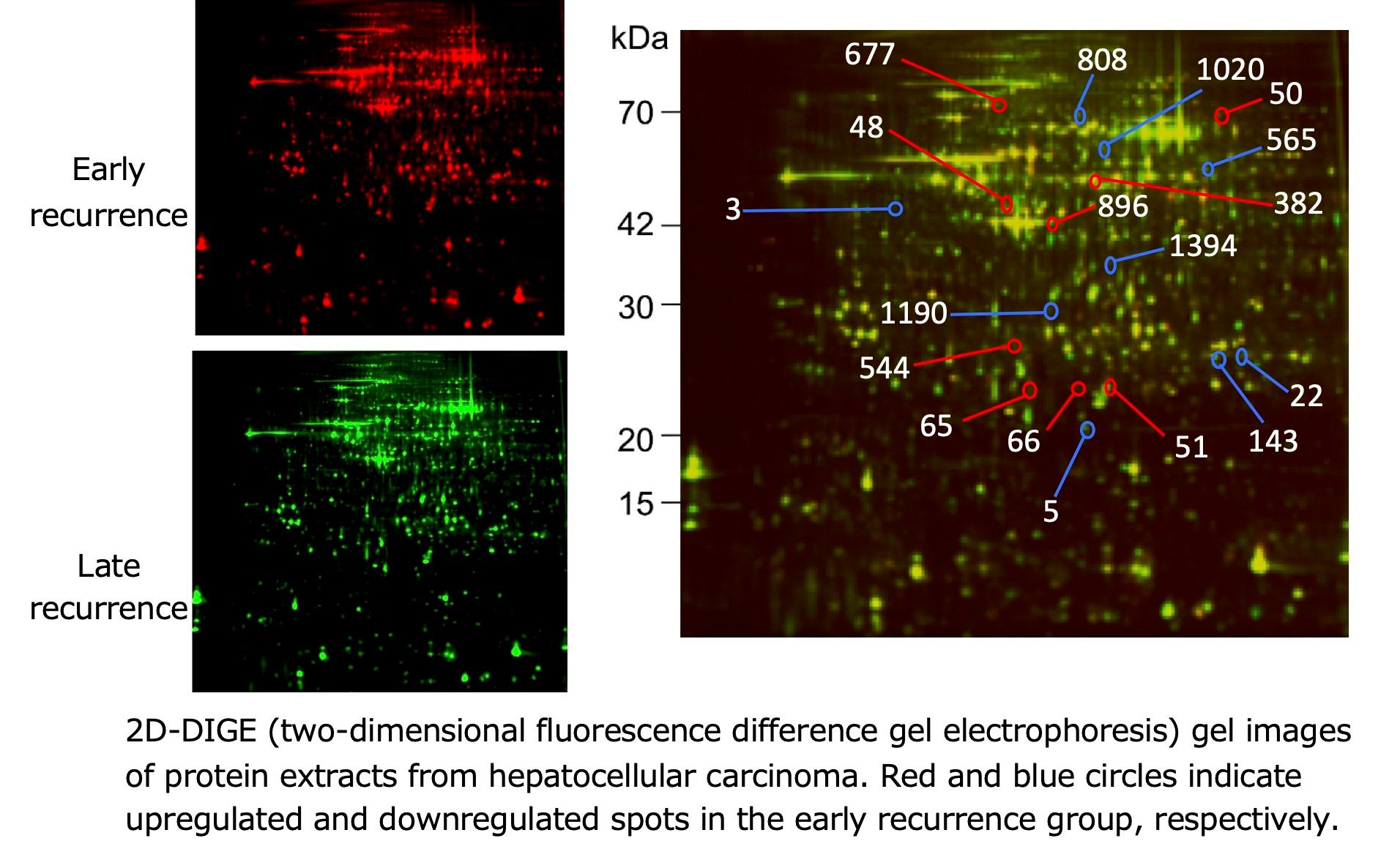
Hepatocellular carcinoma (HCC) recurrence arises from multicentric carcinogenesis or intrahepatic metastasis. Proteomics analysis of two groups of primary HCCs followed by early and late recurrence revealed several proteins associated with early recurrence, probably in the intrahepatic metastasis (Yamaguchi et al., Int J Oncol 2017). Transcriptomics and proteomics of nontumor liver from the above two groups of primary HCCs revealed that several nonparenchyma-derived genes and proteins, microenvironmental factors, were associated with late recurrence, probably in the multicentric carcinogenesis.
Disease omics using laser capture microdissection
Molecular events in the local region specific for disease are useful to elucidate the disease mechanism. These molecular changes are obtained from genomics and proteomics of disease-associated microscopic regions using laser capture microdissection, herein referred to microgenomics and microproteomics. Formalin-fixed paraffin-embedded (FFPE) tissue specimens used for the pathological diagnosis are usable to these omics analyses, leading to the development of molecular diagnosis and molecular target therapy.
- For example:
- An antiviral protein was found to be upregulated in the periportal hepatocytes of HCV-high liver by microproteomics. The protein-knockout cells prepared by gene-editing were sensitive to HCV infection, and the overexpressing cells were resistant to HCV infection in vitro.
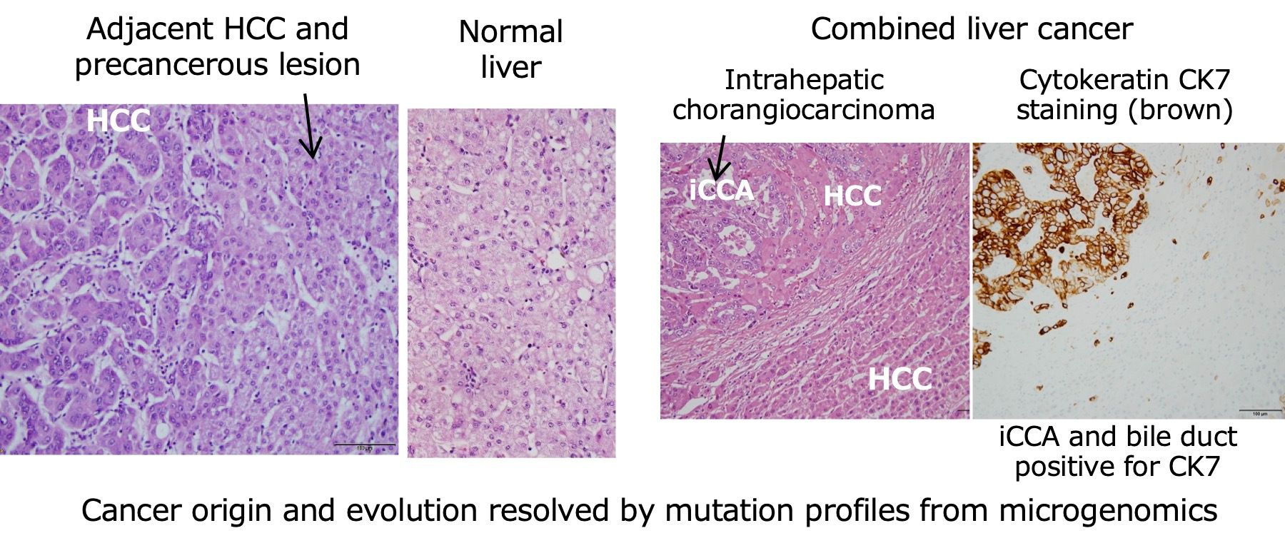
- Cancer origin and evolution have been clarified through somatic mutation profiles by microgenomics of multiple cancer and precancerous lesions: for example, early HCC and precancerous lesion, combined ductal and lobular carcinoma of the breast (Kobayashi et al., Mol Med Rep 2021), tongue cancer and precancerous lesion, and rat HCC, its intrahepatic and lung metastasis.
- Transdifferentiation from HCC to intrahepatic cholangiocarcinoma (iCCA) is a possible path of pathogenesis in combined hepatocellular-cholangiocarcinoma. Both multicentric carcinogenesis and intrahepatic metastasis are involved in three metachronous HCCs every three years. The nontumorous, noncirrhotic liver has a preneoplastic region with a cancer driver mutation in the TERT promoter. (Ohni et al., Oncol Lett 2022)
- Optimization of FFPE fixation and DNA extraction methods is developed for the next generation sequencing (Einaga et al., PLoS ONE 2017). Accurate quantification of DNA including FFPE DNA is qPCR quantification rather than fluorescent dye method (Nakayama et al., PLoS ONE 2016).
Omics of intervertebral disc degeneration
Low-back pain is caused, to some extent, by intervertebral disc degeneration (IVD). One of its etiologic factors is cigarette smoking. The causal relationship and mechanism of degeneration are not well known in vivo. Omics research has been applied to clarify the molecular events in IVD using passive smoking rats.
- IVD degeneration was induced by passive cigarette smoking in rats and was associated with chondrocyte apoptosis and reduction of the extracellular matrix within the IVD (Nakahashi et al., PLoS ONE 2019). Passive cigarette smoking also induced changes in the circadian rhythm of clock genes with a phase shift of 6 hours in rat IVD (Numaguchi et al., J Orthop Res 2016).
- Aging is another etiologic factor in human lumbar disc degeneration. The degeneration level is dependent on the position of lumber disc within a patient. Microproteomics of various levels of disc degeneration revealed several proteins associated with degeneration.
Omics of renal cell carcinoma and head & neck cancer
- VHL gene is a causative of von Hippel-Lindau disease and a tumor suppressor gene. A novel assay for allelic loss of the VHL regardless of heterozygosity was developed (Mochida et al., J Urol 2008). Biallelic inactivation of VHL allelic loss and somatic mutation was unexpectedly low (Hamano et al., J Urol 2002; Igarashi et al., Cancer 2002). VHL allelic loss was also frequently found in tongue cancer (Asakawa et al., Cancer 2008). VHL immunohistochemistry using clinical diagnostic FFPE tissue sections was established and found to be a possible diagnostic marker for precancerous lesion of tongue cancer.
- Maxillary cancer is commonly treated with cisplatin chemotherapy combined with radiotherapy. Tumor protein p53 (TP53) mutation in the carcinoma was found to be more treatment-resistant compared with the carcinoma without TP53 mutation (Kudo et al., Oncol Rep 2010). Transcriptomics of these tumors suggests unexpectedly that TP53 mutated tumors possess a phenotype opposite to that associated with cancer progression and malignant transformation, and exhibit tumor cell heterogeneity between the tumor interior and margins (Kudo et al., Oncol Lett 2017).
- Please also see related references by Dr. Esumi.
We have other research subjects. Please see our publication list.

